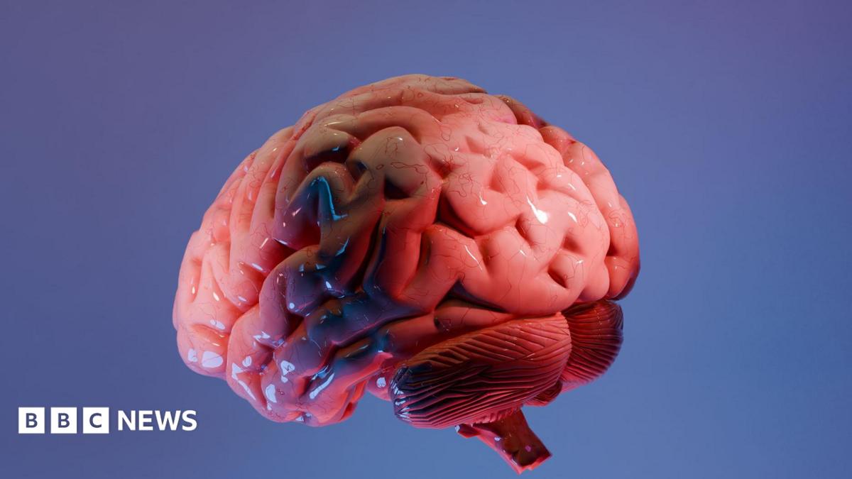
Indian researchers have released a new brainstem atlas, described in media coverage as an unprecedented map of one of the brain’s least known regions. The brainstem atlas is presented as a high-resolution anatomical reference intended to help scientists localise small structures and pathways in this compact, critical part of the central nervous system.
The BBC News report that forms the basis of this article describes fresh anatomical detail and refined boundaries in the brainstem. This explainer summarises what that coverage says, flags where public information is missing, and outlines how a widely available atlas might be used by research groups — while noting that the evaluative term “unprecedented” is a media characterisation of the reported level of detail and should be treated cautiously until methods and validation are publicly available.
What the brainstem atlas shows
According to the BBC account, the atlas brings finer-scale imaging and mapping to the brainstem than appears in many existing references. The coverage reports that the map delineates subdivisions, nuclei and pathways in ways that could help researchers identify tiny anatomical landmarks that are otherwise hard to see with standard neuroimaging.

The article frames the atlas as a shared reference for researchers studying structure, connectivity and disease in the brainstem. While the BBC uses the word “unprecedented” to describe the atlas’ resolution or scope, that is a reporting judgment; without open methods and independent validation, readers should view that claim as provisional rather than definitively established.
Why the brainstem matters
The brainstem connects the brain with the spinal cord and houses circuits that control breathing, heart rate, sleep-wake cycles and basic arousal. It contains many small nuclei and crossing fibre tracts packed into a limited volume, so even modest improvements in anatomical resolution can change how scientists localise functions and interpret clinical findings.
Despite its essential roles, the brainstem has been described as one of the brain’s least known regions because its small, complex structures are difficult to resolve reliably in living humans and because conventional atlases have focused more on cortex and larger subcortical areas. That historical gap is the motivation behind efforts to improve anatomical mapping here.
How the atlas was made and its limits
The BBC report summarises high-level methods such as high-resolution imaging and anatomical mapping techniques but does not publish full technical details. The public account does not name individual researchers or list institutional affiliations in a way that would let readers trace the project to specific labs or publications.
Crucially, the coverage does not link to raw data, processing pipelines, registration parameters, sample sizes or peer-reviewed papers. Those elements are commonly required for independent evaluation: for example, information on how scans were acquired, how images were aligned to a template, what thresholds or statistical checks were used, and how labels were validated against independent samples.
Because those methodological details are absent from the public report, the atlas’ boundaries, labels and generalisability cannot be independently confirmed. The report’s use of the word “unprecedented” is therefore an evaluative description rooted in reporting; it should be read as the BBC’s characterisation of the material presented to them rather than as a settled scientific finding supported by publicly available, peer-reviewed evidence.
What the atlas could enable for scientists
If the atlas is released with open data and thorough documentation, it could improve how scientists map function to structure in the brainstem. More precise anatomical labels can help neuroimaging studies localise small nuclei, make it easier to compare results across studies, and inform targets for neuromodulation or surgical planning where the brainstem is implicated.
Researchers studying sleep disorders, autonomic dysfunction, certain movement disorders and brainstem-mediated reflexes could use a detailed atlas to form more specific hypotheses about which subregions or pathways are involved. Integrating high-resolution anatomical maps with connectivity and histological data could also support multimodal atlases that better link microstructure to function.
However, the extent of these benefits depends on release practices: open access to the atlas files, clear versioning, and validation across different scanners and populations will determine whether the atlas becomes a widely trusted resource or remains a promising but limited reference.
Methods, transparency and next steps
For the atlas to support reproducible research, the following transparency steps would be important: public release of the raw and processed imaging datasets, publication of the registration and labelling pipelines (including parameter settings), disclosure of sample characteristics and counts, and peer-reviewed validation against independent cohorts or histological references.
Sharing data and code would allow other groups to test whether the atlas generalises across different scanners, populations and imaging conditions. Without those elements, scientists can use the atlas as a provisional guide, but independent verification will be needed before it can serve as a definitive resource.
What comes next
Researchers and journals commonly follow up preliminary atlas releases with peer-reviewed papers, open data repositories and collaborative validation studies. If the creators of this atlas publish methods and datasets, the community can begin reproducibility checks, compare the atlas with other references, and explore practical uses in clinical research and neuroimaging workflows.
Absent such material, the immediate next steps for the field will likely be independent attempts to replicate the reported mappings using available datasets and to test how well the atlas labels correspond to functional or clinical markers across different groups.
FAQ
What is a brainstem atlas?
A brainstem atlas is a detailed anatomical map that labels subdivisions, nuclei and major pathways within the brainstem to provide a common reference for researchers and clinicians.
How will this atlas help scientists?
It could help localise function and pathology more precisely, refine targets for neuromodulation, and improve cross-study comparisons — but those benefits depend on transparent release and validation of the underlying data.
Does the report name the researchers or institutions behind the atlas?
The BBC News report summarised the project but did not provide named researchers or detailed institutional affiliations in the public article. That lack of attribution and of shared data or methods is an important limitation for independent verification.
Source attribution: This explainer is based on BBC News coverage of the brainstem atlas. Original coverage is available at https://www.bbc.co.uk/news/articles/cg53l737v1qo. Where the BBC report did not disclose methods or named authors, this article has flagged those gaps and advised caution about evaluative claims such as “unprecedented.”